It is an extraoral radiography method that provides a comprehensive two-dimensional image of the teeth and the skeletal structures of the maxillofacial region. This imaging technique has various applications in dentistry, enabling dentists to accurately diagnose and plan treatments for different oral and dental conditions. In this article, we will discuss the indications for performing panoramic dental imaging and its significance in improving patient care.
Indications for Panoramic Dental Imaging Evaluation of Periodontal Disease: Panoramic dental imaging is useful for assessing periodontal disease and determining the extent of bone defects related to periodontal conditions. It allows for a simultaneous examination of all teeth, assisting in the diagnosis and treatment planning for periodontal conditions.
Orthodontic Assessment: Panoramic dental imaging plays a crucial role in planning orthodontic treatments. It provides information about the presence of tooth germs, the developmental stages of dentition, and the presence of impacted, supernumerary, or retained teeth. These details contribute to the formulation of an effective orthodontic treatment plan.
Assessment of Impacted Third Molars: Panoramic dental imaging is commonly used to evaluate impacted third molars, also known as wisdom teeth. It helps determine the position, angulation, and proximity of the impacted tooth to adjacent structures, aiding in treatment decisions such as extraction or referral to an oral surgeon.
Dental Age Estimation: Panoramic dental imaging is utilized for estimating dental age, particularly in pediatric dentistry and forensic odontology. Dental age assessment is essential for evaluating growth and development, orthodontic treatment planning, and determining the chronological age of an individual in forensic investigations.
Identification of Lesions and Pathologies: Panoramic dental imaging assists in identifying and evaluating various lesions such as tumors, cysts, and other bone diseases that cannot be fully imaged by periapical radiographs. It provides a comprehensive view of the maxillofacial region, enabling prompt diagnosis and appropriate management of these conditions.
Evaluation of Mandibular Trauma: In cases of mandibular trauma, panoramic dental imaging can provide vital information regarding fractures, dislocations, or other traumatic injuries. It aids in the accurate diagnosis and planning of appropriate treatment interventions.
Implant Planning: Panoramic dental imaging is crucial in implant dentistry for treatment planning. It provides detailed information about bone quantity, quality, and anatomical structures, helping determine the optimal location for implant placement.
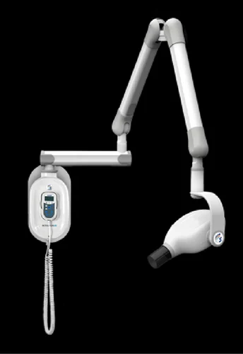
Procedure and Equipment During a panoramic dental imaging procedure, patients are positioned properly using a chin support and bite piece, and a lead apron is used for radiation protection. The imaging machine rotates around the patient's head, capturing a curved image of the teeth, mandible, parts of the maxilla, maxillary sinuses, hard palate, and temporomandibular joints (TMJs). The procedure is safe and relatively comfortable for the patient, requiring little to no special preparation.
Benefits and Limitations Panoramic dental imaging offers several advantages in dental practice. It provides a comprehensive overview of the maxillofacial region, aiding in the accurate diagnosis and treatment planning for various dental conditions. Compared to intraoral X-rays, panoramic images cover a wider area, providing valuable information about tooth positioning, bone abnormalities, sinuses, and impacted teeth. They are commonly used for treatment planning in orthodontics, dentures, extractions, and implants.
However, it's important to note that panoramic dental imaging has certain limitations. Due to its two-dimensional nature, it may not provide detailed information about specific structures or subtle pathologies. In some cases, additional imaging modalities such as cone beam computed tomography (CBCT) or intraoral X-rays.
Provide our customers and partners with an extensive and expert team of clinical, technical, and marketing resources.

Branches
Get in touch
 +91 805 093 1561 or 080 4224 2929
