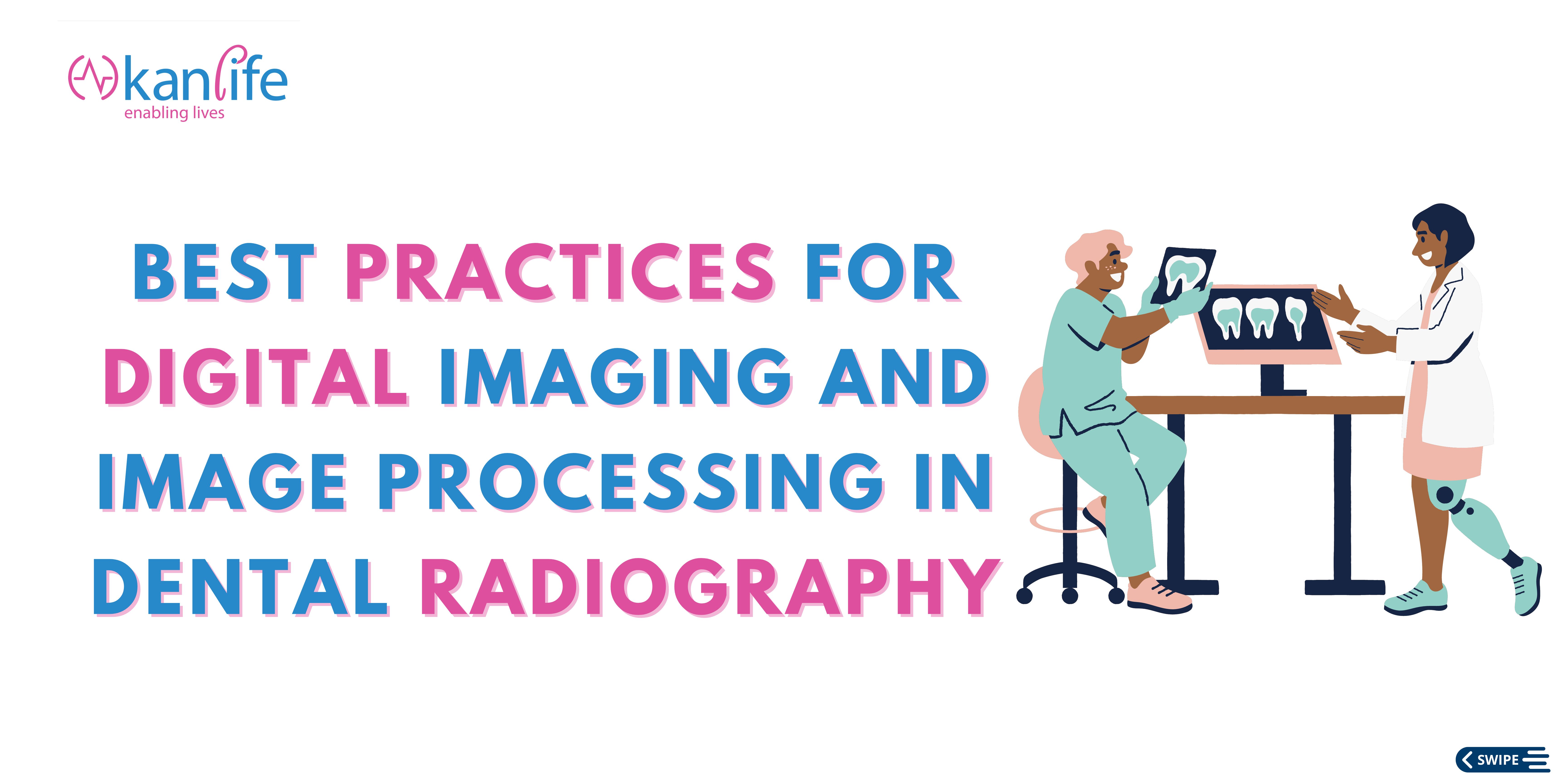
These advancements have allowed dental professionals to capture high-quality images, enhance them for better diagnosis, and improve overall patient care. However, ensuring accurate and reliable results requires following best practices to overcome the challenges associated with digital imaging and image processing in dental radiography. In this article, we will explore these challenges and provide effective solutions.
One of the primary challenges in digital imaging and image processing in dental radiography is obtaining high-quality images with optimal contrast, brightness, and resolution. Low-quality radiographic images can hinder accurate diagnosis and affect treatment evaluation. Factors such as low X-ray radiation levels, limited tool capabilities, and noise presence contribute to the suboptimal quality of radiographic images.
To overcome these challenges, dental professionals can implement the following best practices:
Utilize Direct Digital Imaging: Direct digital imaging techniques, such as solid-state sensors like CCD or CMOS-based chips, offer superior image quality compared to indirect methods. These sensors capture the image directly, eliminating the need for additional scanning or conversion steps. By using direct digital imaging, dental professionals can ensure sharper and more accurate radiographic images.
Optimize Image Enhancement: Image enhancement techniques play a vital role in improving the quality and diagnostic efficacy of dental radiographic images. Dentists can employ various methods, including contrast stretching (CS), contrast limited adaptive histogram equalization (CLAH E), contour let transform (CT), and wavelet transform, to enhance contrast, reduce noise, and optimize brightness. These image enhancement techniques can help in better evaluating the condition of dental structures such as fillings, pulp tissue, and periodontal ligaments.
Implement Advanced Software and Tools: Dental imaging software and tools provide extensive capabilities for image processing and analysis. These tools allow dental professionals to manipulate images, apply filters for detecting carious lesions, and visualize supporting tissues with high resolution. By utilizing advanced software, dentists can improve the accuracy of diagnosis and treatment evaluation.
Stay Updated with Technology: The field of dental radiography is constantly evolving, with new technologies and techniques emerging regularly. Dental professionals must stay updated with the latest advancements and incorporate them into their practices. Regularly attending conferences, workshops, and continuing education programs can help dentists enhance their skills in digital imaging and image processing. By staying updated with technology, dental professionals can ensure they are utilizing the most effective tools and techniques available.
In conclusion, digital imaging and image processing have significantly transformed dental radiography, enabling precise diagnosis and effective treatment planning. By following best practices such as utilizing direct digital imaging, optimizing image enhancement, implementing advanced software and tools, and staying updated with technology, dental professionals can ensure the production of high-quality radiographic images and enhance patient care. Embracing these best practices will not only improve the accuracy and efficiency of dental radiography but also contribute to better treatment outcomes and patient satisfaction.
Skanray Dental X-ray is a valuable solution that aligns perfectly with the challenges and best practices discussed in the article. Skanray offers advanced dental imaging equipment and technologies that can greatly enhance digital imaging and image processing capabilities in dental radiography. By incorporating Skanray Dental X-ray into their practices, dental professionals can benefit from its features and contribute to improved patient care.
One of the primary challenges in dental radiography is obtaining high-quality images with optimal contrast, brightness, and resolution. Skanray Dental X-ray addresses this challenge by providing direct digital imaging techniques. With solid-state sensors like CCD or CMOS-based chips, Skanray Dental X-ray captures images directly, eliminating the need for additional scanning or conversion steps. This ensures sharper and more accurate radiographic images, enabling precise diagnosis and effective treatment planning.
To optimize image enhancement, Skanray Dental X-ray offers advanced image processing capabilities through its software and tools. Dental professionals can manipulate images, apply filters for detecting carious lesions, and visualize supporting tissues with high resolution. By utilizing the advanced software provided by Skanray, dentists can enhance the quality and diagnostic efficacy of dental radiographic images, thus improving the accuracy of diagnosis and treatment evaluation.
Moreover, Skanray understands the importance of staying updated with technology in the field of dental radiography. They constantly strive to aggregate the best-in-world technologies, partnering with world-renowned organizations for designing and manufacturing their equipment. By choosing Skanray Dental X-ray, dental professionals gain access to cutting-edge technologies and stay at the forefront of advancements in digital imaging and image processing.
Skanray Dental X-ray is a valuable asset for dental professionals looking to overcome the challenges associated with digital imaging and image processing in dental radiography. With its direct digital imaging techniques, advanced image enhancement capabilities, and commitment to staying updated with technology, Skanray Dental X-ray empowers dental professionals to produce high-quality radiographic images, enhance patient care, and contribute to better treatment outcomes and patient satisfaction.
Say Goodbye to Suboptimal Images: Overcome the challenges of low-quality radiographic images with Skanray Dental X-ray's direct digital imaging techniques, delivering sharper and more accurate results.
Provide our customers and partners with an extensive and expert team of clinical, technical, and marketing resources.

Branches
Get in touch
 +91 805 093 1561 or 080 4224 2929
